
IN-2026-018 - Common Ivy (Hedera helix) — Leaf (W.M.), Partial Clearing
Specimen & Context
| Date | 2026-04-15 |
| Species | Hedera helix |
| Common Name | Common Ivy |
| Habit | Climbing plant, capable of both flexible extension and longer-term structural support |
| Material | Fresh leaf |
| Location | Abingdon, Oxfordshire, UK |
| Preparation | Leaf, Whole Mount (W.M.), clearing with household materials |
| Stain | None |
| Series | Scheme of Structural Investigations - Series II — Support & Conduction |
Overview
This investigation set out to examine the venation of a leaf of Hedera helix by means of chemical clearing, with the aim of rendering the vascular structure visible in whole mount.
Clearing was attempted using readily available household reagents. While full transparency of the lamina was not achieved, the process yielded a series of partially cleared preparations in which epidermal, mesophyll, and vascular elements could be observed in varying degrees.
Particular attention was given to the effect of preparation on visibility of structure, and to the role of tissue permeability in determining the outcome.
Method (Summary)
- Fresh leaf material selected (including both thicker and thinner samples)
- Specimens subjected to repeated cycles of:
- Dilute sodium hypochlorite (bleach)
- Hot sodium bicarbonate solution
- Intermediate rinsing in water between treatments
- Selected areas mechanically disturbed (scraping/teasing) to aid penetration
- Sections mounted in water (W.M.) under cover glass
- Observed under moderate magnification across multiple fields
Observations
- Full clearing of the lamina was not achieved in either specimen
- Marked differences were observed between thicker and thinner leaf material:
- Thicker leaf remained largely bright green and opaque
- Thinner leaf became partially cleared, with brownish discolouration
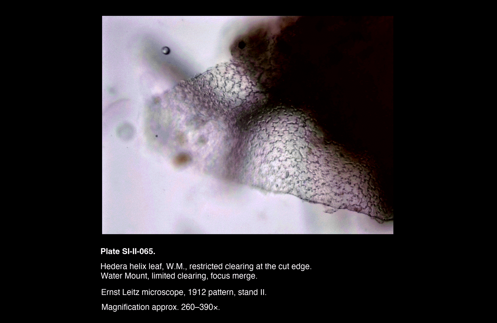
- Clearing was most evident at cut edges, where:
- Tissue became more translucent
- Underlying structure was more readily resolved
- In partially cleared regions:
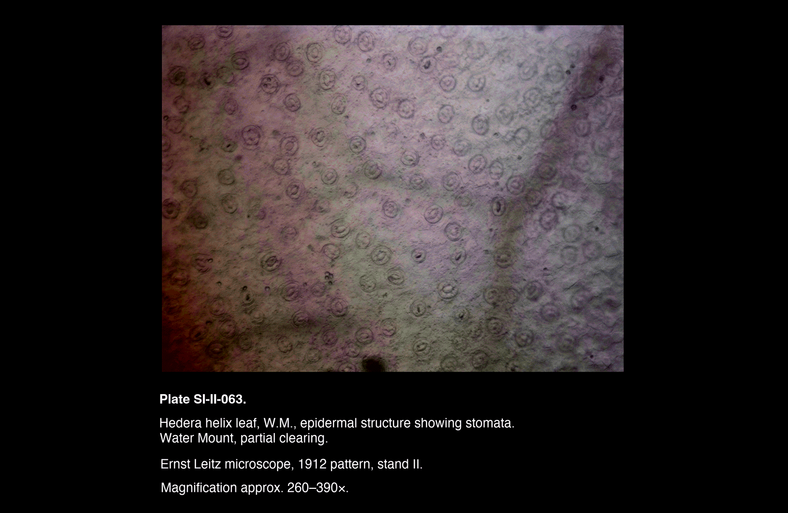
- Stomata were clearly visible as numerous circular or oval structures across the epidermis
- Mesophyll appeared as a mottled, partially degraded matrix
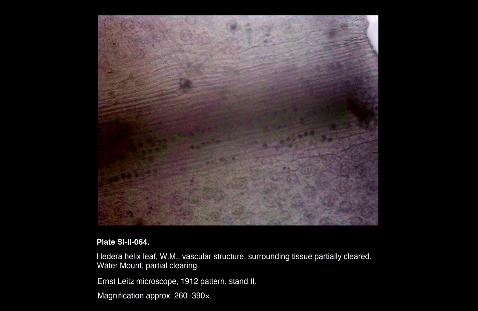
- Veins were present but often indistinct, appearing as faint linear structures beneath remaining tissue
- Major veins were most readily observed:
- Near cut edges
- Where local clearing was greatest
- Focusing was often difficult due to:
- Residual tissue thickness
- Overlapping structural layers within the preparation
Plates
Selected Plates
/Hedera%20helix%20leaf,%20W.M.,%20epidermal%20structure%20showing%20stomata.)
/Hedera%20helix%20leaf,%20W.M.,%20vascular%20structure,%20surrounding%20tissue%20partially%20cleared.)
/Hedera%20helix%20leaf,%20W.M.,%20restricted%20clearing%20at%20the%20cut%20edge)
Plates
Selected Plates
Interpretation
Effectiveness of Clearing
The clearing process was only partially effective. While some pigment removal and tissue softening occurred, the lamina was not rendered sufficiently transparent to isolate the venation.
This appears to reflect a combination of:
- Limited chemical strength of the reagents used
- Incomplete penetration through the intact cuticle and epidermis
- Structural robustness of the leaf tissue
Clearing was consistently most successful at cut edges, indicating that access to internal tissue is a primary limiting factor.
Visibility of Structure
The partially cleared preparations reveal a layered structure:
- The epidermis, including stomata, is readily observed once some transparency is achieved
- The mesophyll remains optically significant, scattering light and obscuring deeper structures
- The vascular system is present but difficult to resolve except where surrounding tissue is reduced
As a result, visibility of venation is not simply a function of its presence, but of the optical properties of surrounding tissue.
Permeability and Preparation
This investigation highlights the importance of permeability in chemical preparation:
- Intact surfaces limit reagent access
- Mechanical disruption (cutting, scraping) improves local outcomes
- Chemical treatment alone may be insufficient without adequate access
The contrast between cut and uncut regions provides direct evidence of this effect.
Structural Relationships
Despite incomplete clearing, the relationship between tissue types is evident:
- Stomata are confined to the epidermal layer and remain prominent
- Mesophyll forms a continuous but variably degraded matrix
- Veins persist as deeper, more coherent structures, becoming visible where mesophyll is reduced
This layered organisation is preserved even when full isolation of structures is not achieved.
Methodological Note
This investigation demonstrates that chemical clearing is not a binary outcome but a continuum of preparation states:
- Untreated tissue → structurally intact but optically dense
- Partial clearing → increased transparency with multiple layers visible
- Full clearing → isolation of vascular structure (not achieved here)
The present preparations occupy an intermediate position, which, while not fulfilling the original aim, provides a broader view of tissue organisation in situ.
Remarks
- Thinner leaf material responded more readily to treatment than thicker material
- Cut edges consistently provided the most informative views
- Stomatal distribution was particularly well demonstrated in partially cleared preparations
- Complete clearing appears to require either:
- Stronger reagents, or
- More effective disruption of surface barriers
- The investigation yielded useful observations despite not achieving full transparency
- The results emphasise the importance of adapting preparation to both specimen type and available reagents