
IN-2026-011 - Common Daisy (Bellis perennis) — Petal (W.M.)
Specimen & Context
| Date | 2026-03-28 |
| Species | Bellis perennis |
| Common Name | Common Daisy |
| Structure | Floral display structure (ray floret) forming part of the composite flower head |
| Material | Fresh petal |
| Location | Abingdon, Oxfordshire, UK |
| Preparation | Petal, Whole Mount (W.M.) |
| Stain | None |
| Series | Scheme of Structural Investigations - Series III — Reproductive Elements |
Overview
This investigation examines the cellular structure of a petal from Bellis perennis (Common Daisy) using a whole mount preparation. The aim was to observe epidermal organisation, cell morphology, and structural variation across the petal surface, with particular attention to changes at the margin.
Method (Summary)
- A small section of petal removed and mounted flat without water
- Cover slip applied carefully to avoid folding or compression
- Observed under medium to high power objectives
- Multiple fields recorded, including petal margin
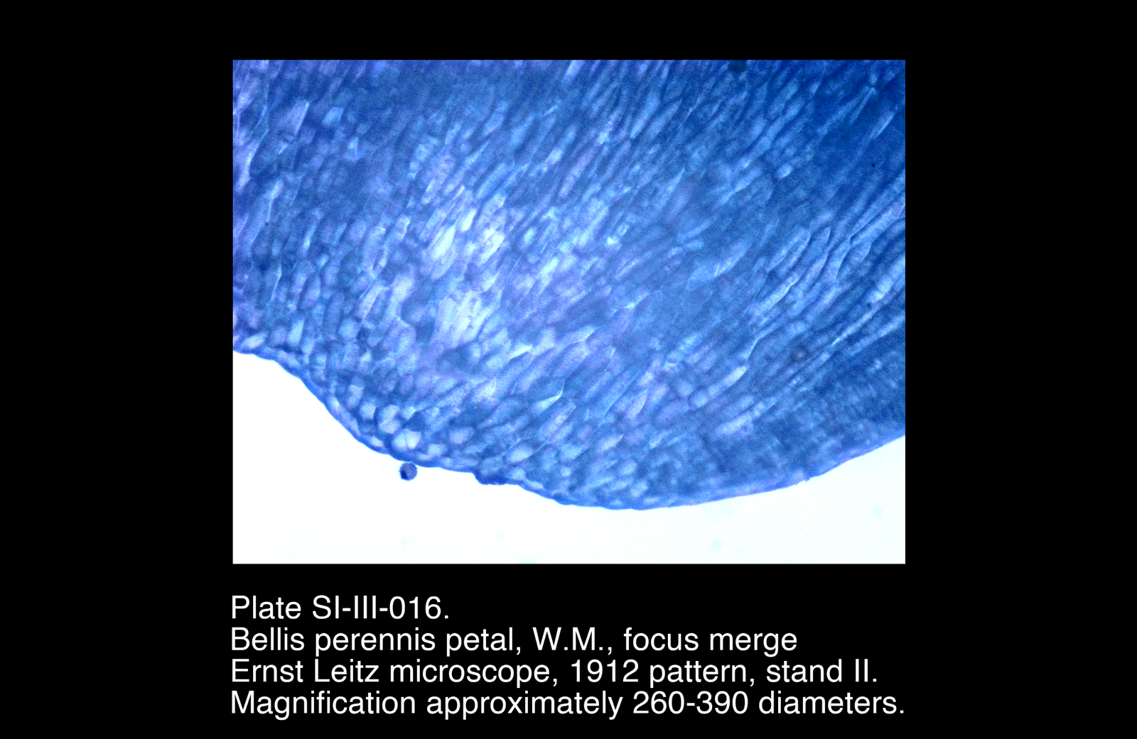
- A focus stack was combined to produce a merged image for improved depth clarity
Observations
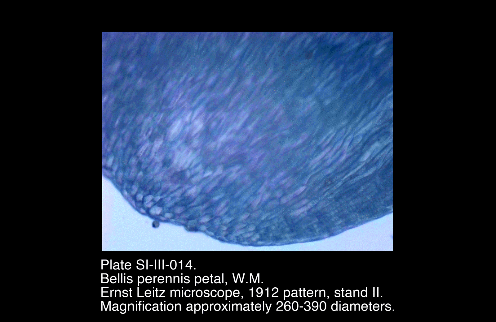
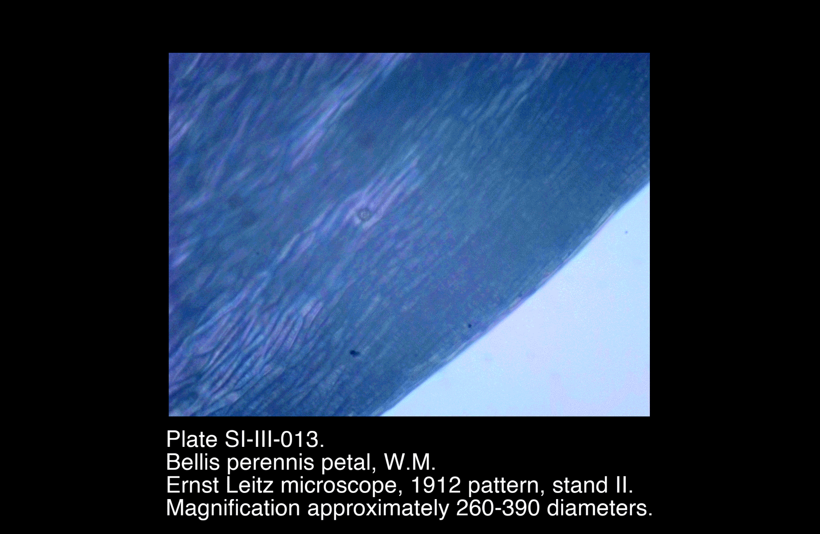
- Surface composed of elongated epidermal cells, aligned longitudinally along the petal axis
- Cell walls clearly defined
- Cells exhibit slight waviness rather than strict rectangular geometry
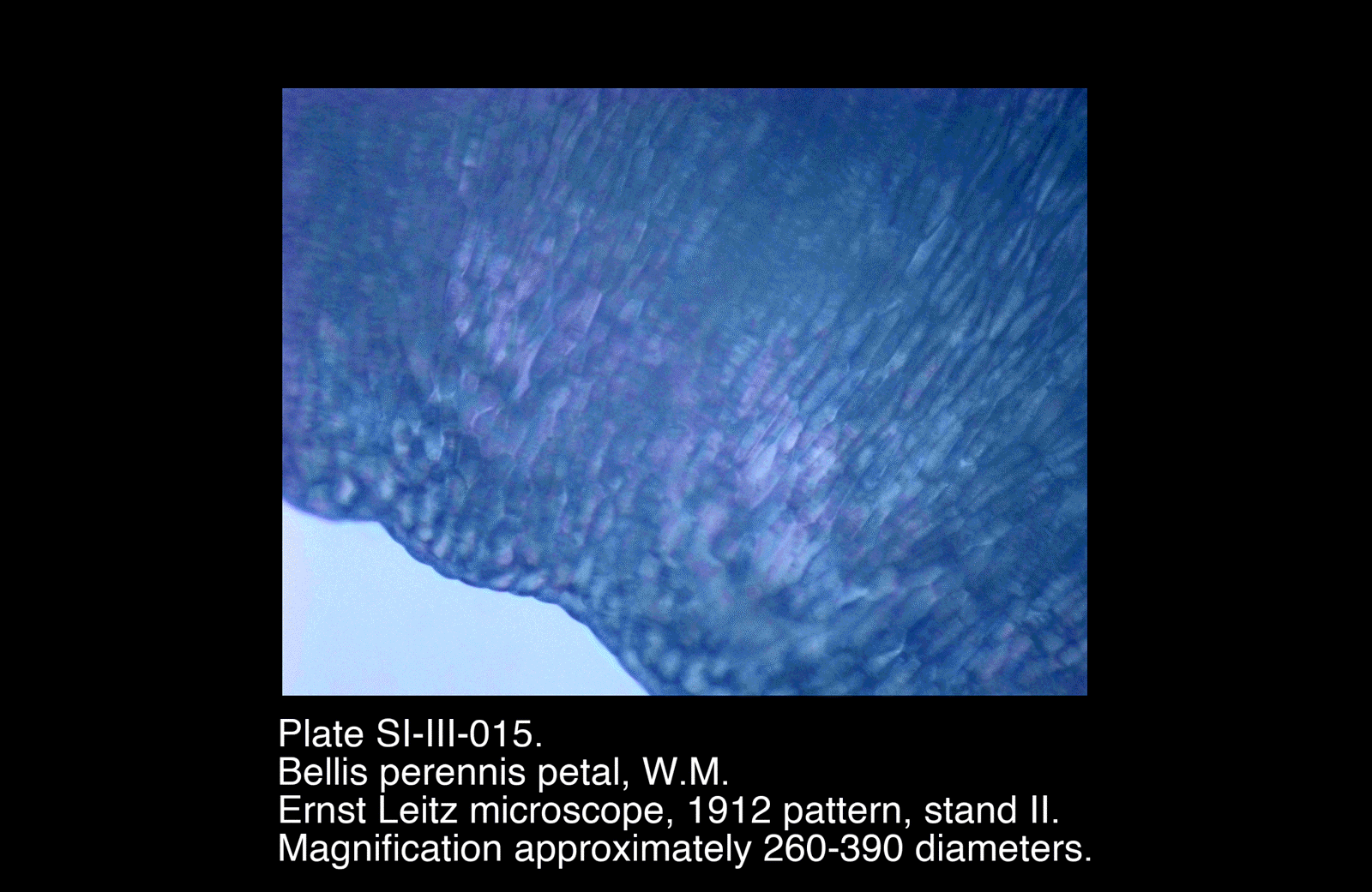
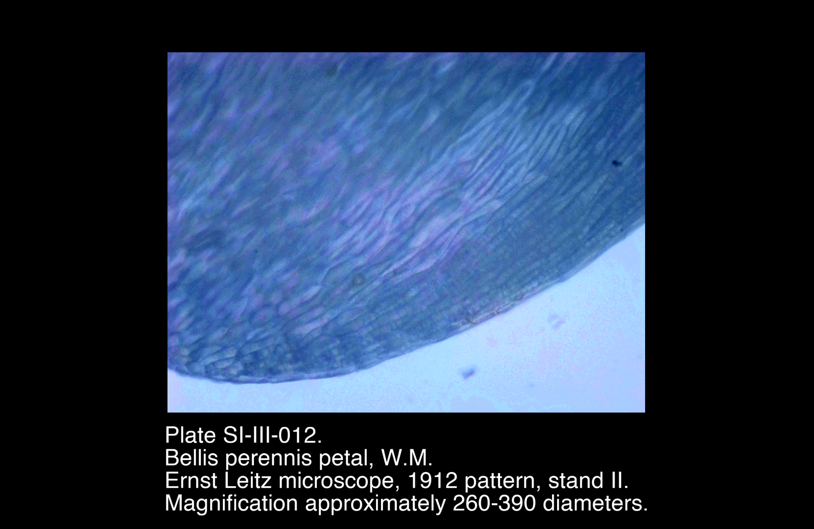
- Toward the petal margin:
- Cells become smaller and more rounded
- Packing becomes denser and less ordered
- No clear stomatal structures observed
- Occasional small spherical bodies present at the margin, not integrated with tissue structure (likely debris or pollen)
- Focus merge reveals layering and depth variation not fully visible in single focal planes
Plates
Selected Plates
/Bellis%20perennis%20petal,%20W.M.)
/Bellis%20perennis%20petal,%20W.M.)
/Bellis%20perennis%20petal,%20W.M.)
Plates
Selected Plates
These plates show the clearest representation of epidermal cell organisation and the transition in morphology toward the petal edge. The focus-merged image provides improved depth resolution across the tissue.
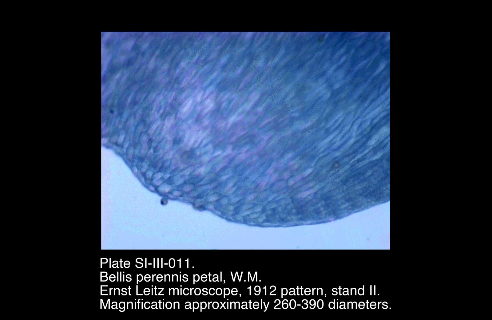
Preliminary Plates
/Bellis%20perennis%20petal,%20W.M.)
/Bellis%20perennis%20petal,%20W.M.)
/Bellis%20perennis%20petal,%20W.M.)
Preliminary Plates
Earlier plates show similar structure but with reduced clarity or depth of focus. These were useful in identifying consistent features and refining mounting and focusing technique.
Interpretation
The petal of Bellis perennis exhibits a highly regular epidermal structure, reflecting its role as a visual display surface rather than a structural or transport organ.
Surface Organisation
The elongated, longitudinally aligned cells indicate directional growth along the petal axis. Their slight waviness suggests expansion during development rather than rigid packing.
The uniformity of this arrangement contributes to a smooth and continuous surface, likely important for optical properties such as light reflection and colour presentation.
Margin Structure
The transition to smaller, more rounded cells at the margin represents a change in growth dynamics at the boundary of the tissue.
This region is:
- More compact
- Less regularly ordered
- Structurally distinct from the main body of the petal
Such differentiation is typical of growth termination zones in plant tissues.
Functional Interpretation
Unlike stems or leaves, the petal shows:
- No evident transport structures
- No stomatal complexes
- Minimal structural reinforcement
Instead, it can be understood as a specialised epidermal sheet, optimised for:
- Visual display
- Surface area
- Light interaction
The simplicity of internal structure reflects this highly specific function.
Remarks
- The whole mount technique proved highly effective for this specimen, preserving natural structure without distortion
- The focus merge significantly improved interpretability, particularly near the margin
- The absence of stomata was notable and consistent with the petal’s function
- This investigation provides a useful contrast to structural tissues examined in earlier studies