
IN-2026-009 - Human Blood — Peripheral Smear (W.M.)
Specimen & Context
| Date | 2026-03-28 |
| Collection Note | Specimen obtained opportunistically following a minor finger cut sustained during field collection; please don't try this deliberately at home! |
| Species | Homo sapiens |
| Common Name | Human |
| Material | Capillary blood (finger) |
| Location | Abingdon, Oxfordshire, UK |
| Preparation | Peripheral blood smear, Wet Mount (W.M.) |
| Stain | None |
| Series | Scheme of Structural Investigations - Series II — Support & Conduction |
Overview
This investigation examines the structure of human blood as a circulating fluid tissue, prepared as a thin peripheral smear and observed without staining. The aim was to explore the morphology and distribution of blood cells, and to contrast this form of conduction with the fixed vascular systems previously examined in plants.
It represents a transition from structural conduction systems (plant vascular tissue) to mobile conduction (animal blood).
Method (Summary)
- A small drop of capillary blood placed near the end of a clean slide
- Spread using a second slide at ~30–45° to produce a thin film
- Multiple smears prepared to improve consistency
- Preparations allowed to air dry
- Observed under high magnification using transmitted light
- Illumination and contrast adjusted to improve visibility of unstained cells
Observations
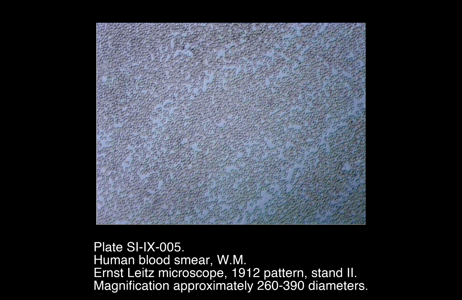
- Field dominated by numerous circular, disc-shaped elements
- Individual cells exhibit:
- Pale central region
- More defined outer margin
- Cells generally uniform in size and shape
- Distribution varies with smear thickness:
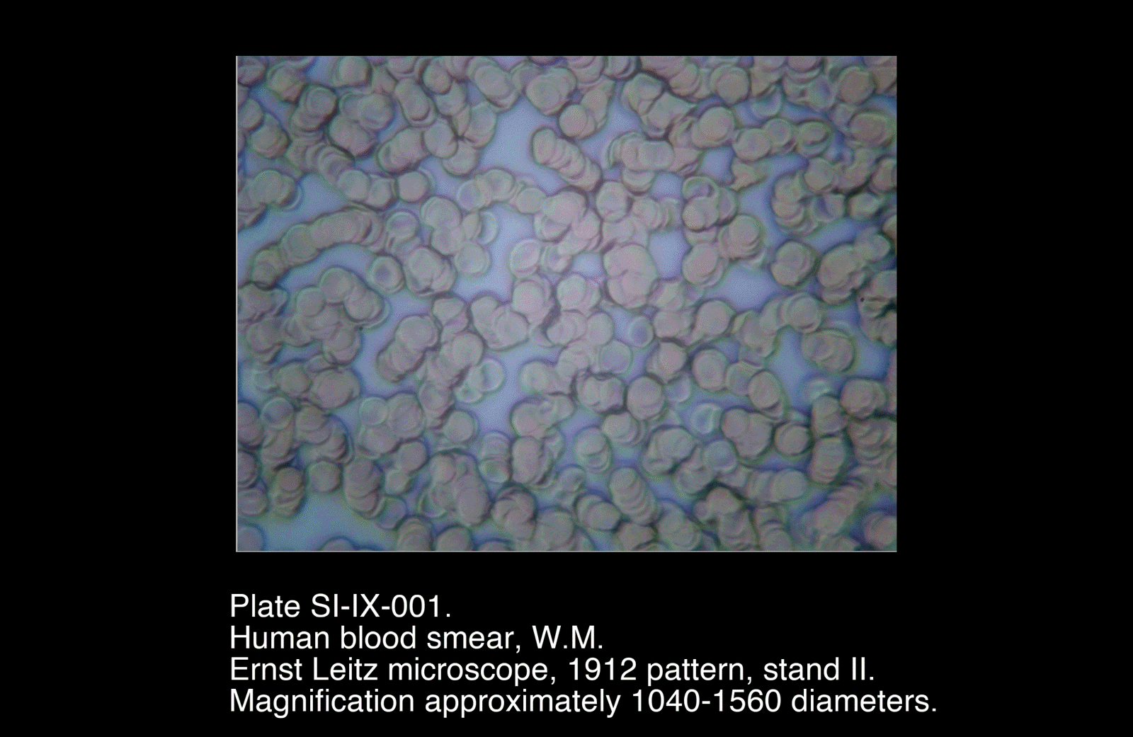
- Thicker regions: cells crowded, overlapping, and distorted
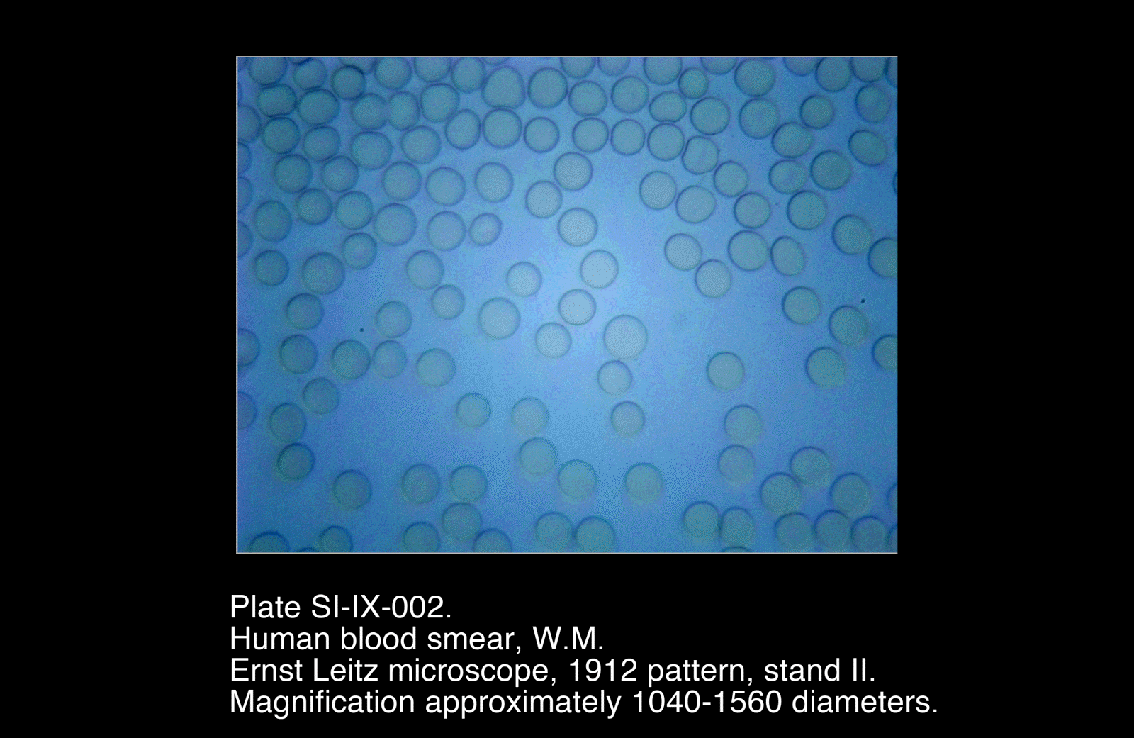
- Thinner regions: cells lie mostly separate and evenly dispersed
- No internal structures clearly visible within individual cells
- Occasional minor irregularities in shape in more crowded areas
Plates
Selected Plates
/Human%20Blood%20Smear,%20W.M.)
/Human%20Blood%20Smear,%20W.M.)
Plates
Selected Plates
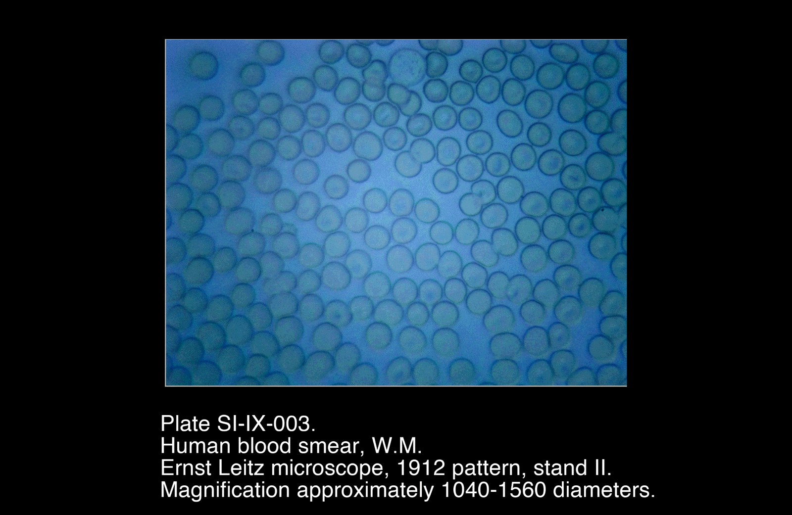
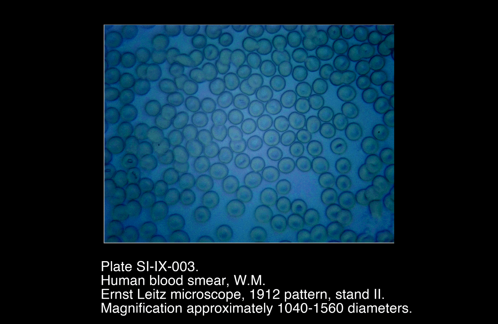
These plates show the most suitable regions of the smear, where cells are sufficiently separated to observe individual morphology.
Preliminary Plates
/Human%20Blood%20Smear,%20W.M.)
/Human%20Blood%20Smear,%20W.M.)
/Human%20Blood%20Smear,%20W.M.)
Preliminary Plates
Earlier preparations include thicker regions of the smear, where overlapping cells reduce clarity but illustrate the effect of preparation technique.
Interpretation
Cell Form
The dominant elements observed are red blood cells (erythrocytes), which appear as biconcave discs. The characteristic pale centre reflects the reduced thickness at the centre of the cell.
The absence of visible internal structure is consistent with the known structure of these cells in mammals, which lack a nucleus in the mature state.
Smear Structure
The smear forms a gradient of thickness, from dense accumulation to a thin, well-dispersed region.
- Thick regions obscure individual cell form
- Optimal observation occurs just behind the feathered edge, where cells are:
- Intact
- Minimally overlapping
- Representative of true morphology
This variation highlights the importance of preparation technique in microscopic interpretation.
Optical Characteristics
In the absence of staining, visibility is governed primarily by:
- Differences in optical thickness
- Edge contrast
- Light scattering at cell boundaries
The central pallor and peripheral definition of each cell are therefore optical effects as much as structural ones.
Functional Interpretation
Blood represents a fundamentally different conduction system from plant vascular tissue:
- Mobile rather than fixed
- Composed of discrete cellular elements suspended in fluid
- Capable of continuous circulation rather than directional flow through defined vessels
The uniformity and abundance of erythrocytes reflect their primary role in gas transport, forming the bulk of the circulating cellular material.
Remarks
- Considerable variation between smears highlights the importance of:
- Drop size
- Spreader angle
- Speed of preparation
- Best results obtained where the smear was thin and evenly distributed
- Unstained preparations provide limited structural detail but clearly demonstrate overall cell form and distribution
- Future work may include stained preparations to reveal additional cellular components (e.g. white blood cells)
This investigation provides a useful contrast to plant-based conduction systems, extending the series into animal transport mechanisms.