
IN-2026-004 - Cypress-leaved Plait-moss (Hypnum cupressiforme) — Whole Mount (W.M.)
Specimen & Context
| Date | 2026-03-18 |
| Species | Hypnum cupressiforme |
| Common Name | Cypress-leaved Plait-moss |
| Habit | Pleurocarpous moss forming low, spreading mats |
| Material | Fresh moss shoot, individual leaves examined |
| Location | Abingdon, Oxfordshire, UK |
| Preparation | Whole Mount (W.M.) |
| Stain | None |
| Series | Scheme of Structural Investigations - Series II — Support and Conduction |
Overview
This investigation examines the leaf structure of Hypnum cupressiforme (Cypress-leaved Plait-moss) by whole mount. The aim was to observe cellular organisation within the leaf (phyllid), and to consider how structure supports exchange and function in a non-vascular plant.
Method (Summary)
- Individual leaves removed from shoot and mounted in water (W.M.)
- Observed under low and high power objectives
- Attention given to both the leaf margin and apex, and to the central region (costa)
- Focus-merge imaging used to improve depth of field across the curved leaf surface
Observations
- Leaves thin, translucent, and lanceolate, tapering to a fine point
- Clear central costa (midrib) visible, extending toward the apex
- Lamina composed of numerous small, regularly arranged cells, giving a fine reticulate appearance
- Cells appear elongate to polygonal, aligned along the length of the leaf
- Leaf margin finely toothed or uneven, particularly near the apex
- No evidence of vascular bundles or differentiated transport tissues
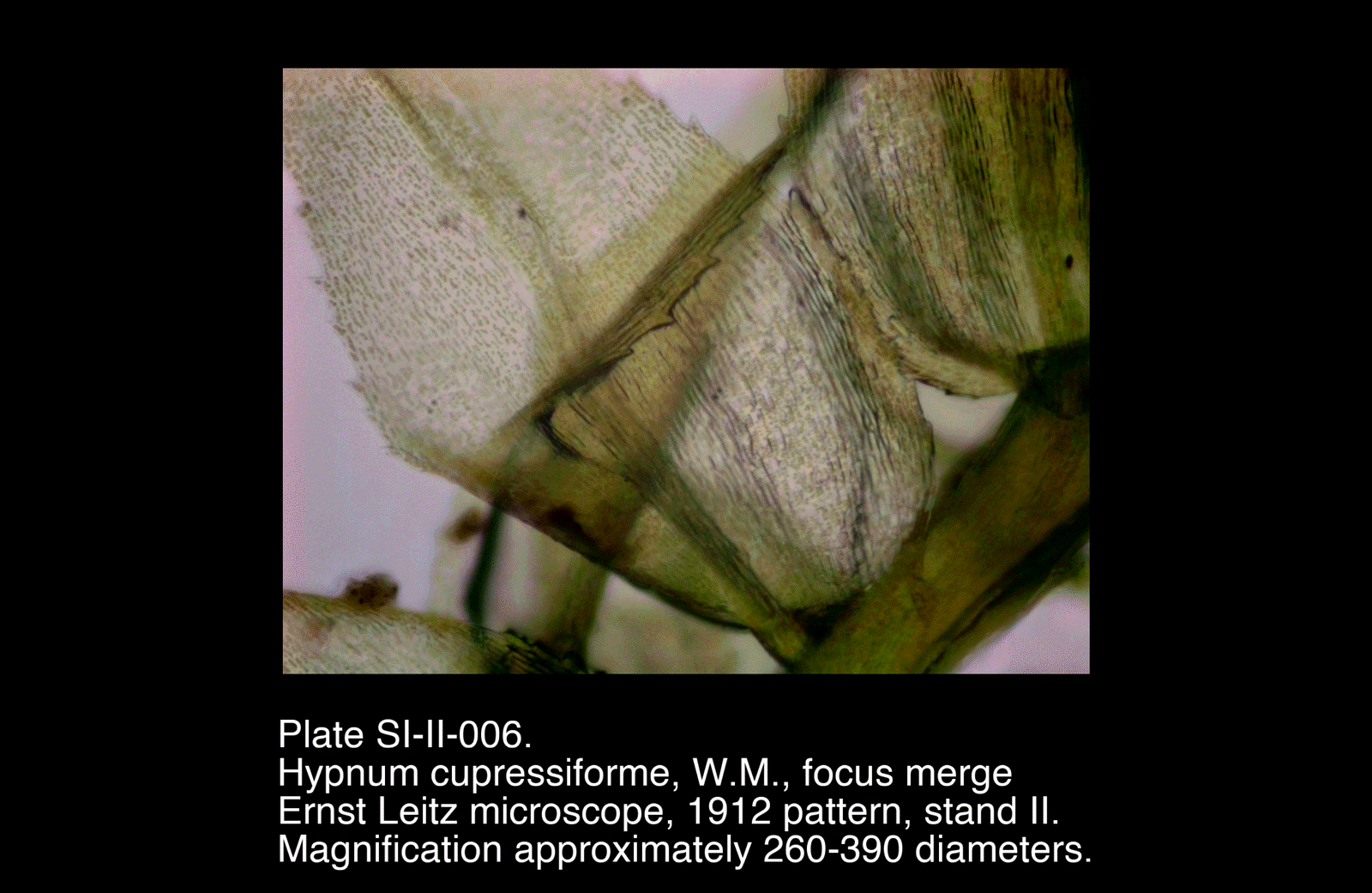
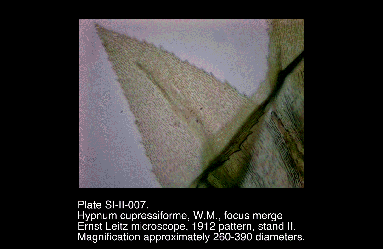
- Focus-merged plates (006–007) significantly improve clarity of cell pattern and costa
Plates
Selected Plates
/Hypnum%20cupressiforme,%20W.M.,%20focus%20merge)
/Hypnum%20cupressiforme,%20W.M.,%20focus%20merge)
Plates
Selected Plates
These plates show the clearest resolution of the cellular pattern within the lamina and the structure of the costa.
Preliminary Plates
/Hypnum%20cupressiforme,%20W.M.)
/Hypnum%20cupressiforme,%20W.M.)
Preliminary Plates
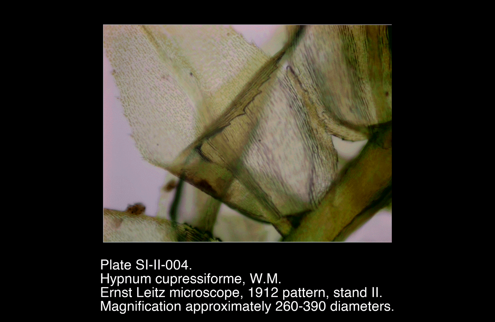
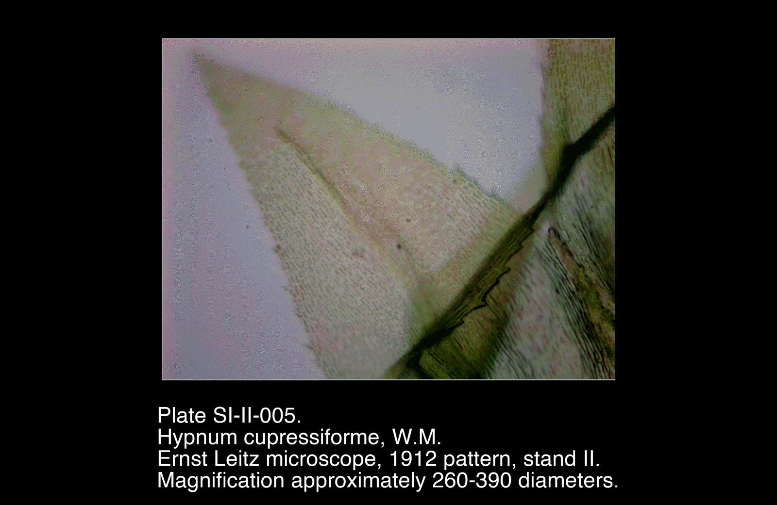
Earlier views establish general leaf form and orientation, though with reduced clarity of fine cellular detail.
Interpretation
The leaf structure of Hypnum cupressiforme represents a simple, non-vascular photosynthetic surface, distinct from both lichen thalli and higher plant leaves.
Surface and Cellular Organisation
The lamina is composed of a single layer of small cells, forming a continuous surface. This arrangement maximises:
- Exposure to light
- Direct exchange with the surrounding environment
- Efficient uptake of water across the entire leaf surface
The regularity of the cell pattern contrasts with the more diffuse internal organisation observed in the lichen thallus.
Costa (Midrib)
The presence of a costa introduces a degree of structural differentiation:
- Provides mechanical support to the otherwise delicate lamina
- May assist in limited conduction of water along the leaf
However, this is not equivalent to the vascular system of higher plants, and remains relatively simple.
Absence of Vascular Tissue
No discrete vascular bundles are present. The leaf relies instead on:
- Surface absorption of water
- Short-distance internal movement between adjacent cells
This reflects the ecological strategy of mosses, which are closely tied to moist environments.
Functional Interpretation
The moss leaf may be understood as a thin, permeable photosynthetic sheet:
- Maximising surface exposure
- Minimising structural investment
- Supported by a simple central reinforcement (costa)
- Functioning without specialised conducting tissues
Comparison within Series I
In relation to Xanthoria parietina:
- Both emphasise surface exposure and exchange
- Moss shows clear cellular organisation, unlike the composite lichen thallus
- Introduction of a costa represents an early step toward structural differentiation
In contrast to vascular plant material, the moss leaf remains entirely dependent on external moisture and lacks internal transport systems.
Remarks
- Focus merging (SI-II-006 and SI-II-007) was particularly effective in resolving the lamina cell pattern
- Careful separation and flattening of leaves improved visibility of structure
- The costa provides a useful reference feature for orientation and interpretation
- A transverse section of the leaf would be a valuable extension of this investigation