
IN-26-003 - Common Orange Lichen (Xanthoria parietina) — Whole Mount (W.M.)
Specimen & Context
| Date | 2026-03-18 |
| Species | Xanthoria parietina |
| Common Name | Common Orange Lichen |
| Habit | Foliose lichen, forming lobed, leaf-like expansions attached to bark, stone, or other exposed surfaces |
| Material | Fresh lichen thallus |
| Location | Abingdon, Oxfordshire, UK |
| Preparation | Whole Mount (W.M.) |
| Stain | None |
| Series | Scheme of Structural Investigations - Series I — Dormant and Elementary Forms |
Overview
This investigation examines the thallus of Xanthoria parietina (Common Orange Lichen) by whole mount. The aim was to observe the general surface structure and internal visual texture of the lichen body, and to consider how its organisation supports exposure, exchange, and persistence on open substrates.
Method (Summary)
- Small portions of thallus mounted whole in water (W.M.)
- Observed under low and high power objectives
- Several views taken to compare thallus margin, broader surface texture, and more detailed internal appearance
- Later plates included focus-merge images to improve depth of field across uneven material
Observations
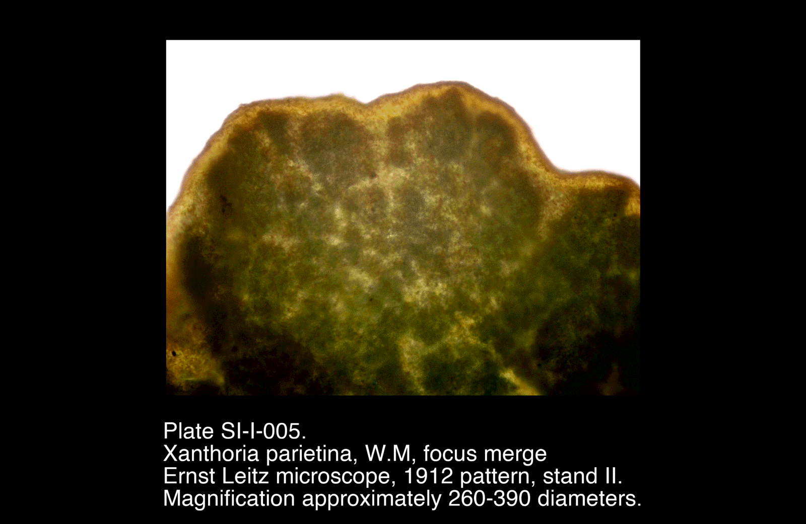
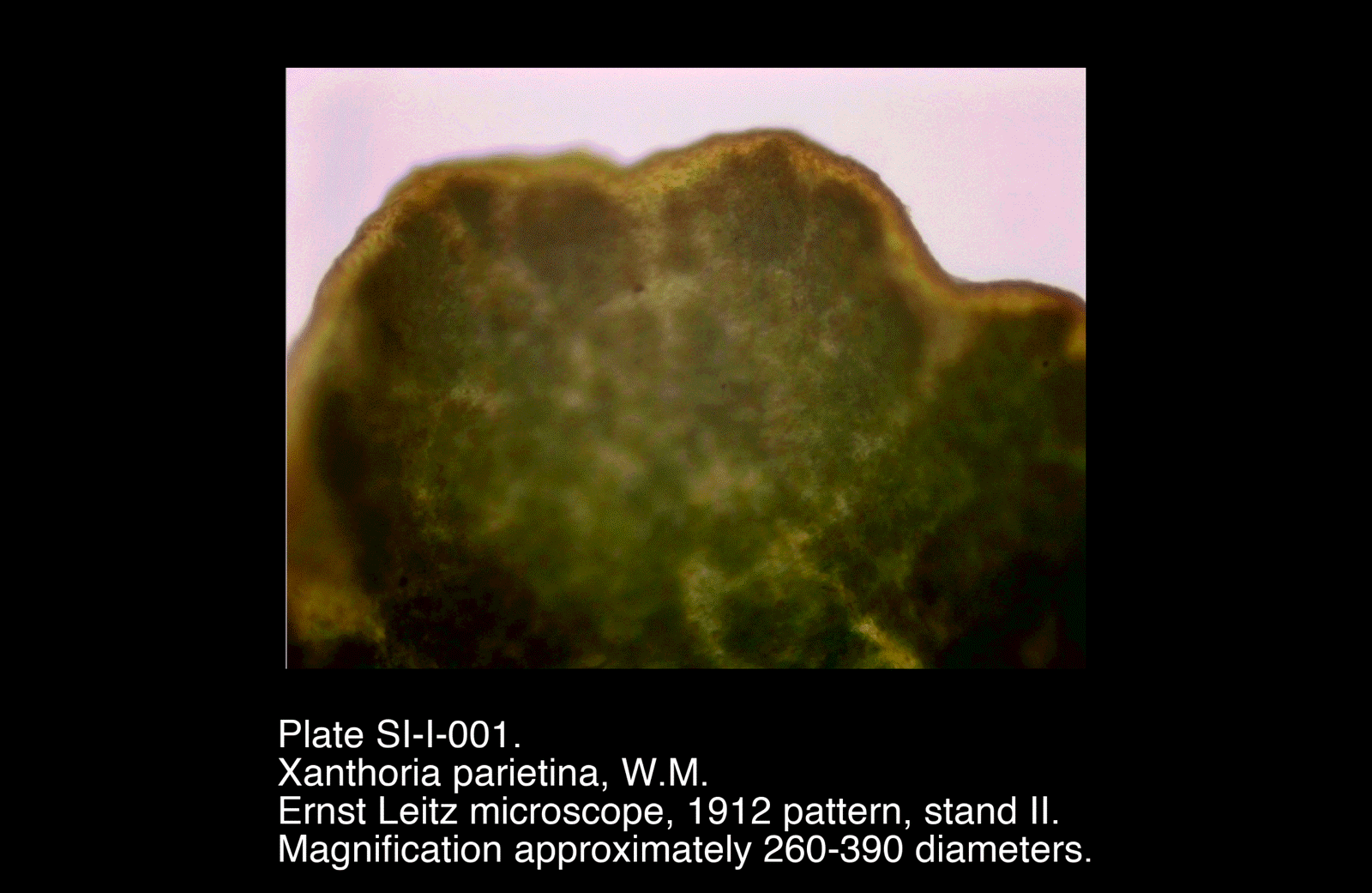
- Thallus margin lobed and undulating, rather than smooth or regular
- Surface appears continuous but uneven, with gentle relief across the upper face
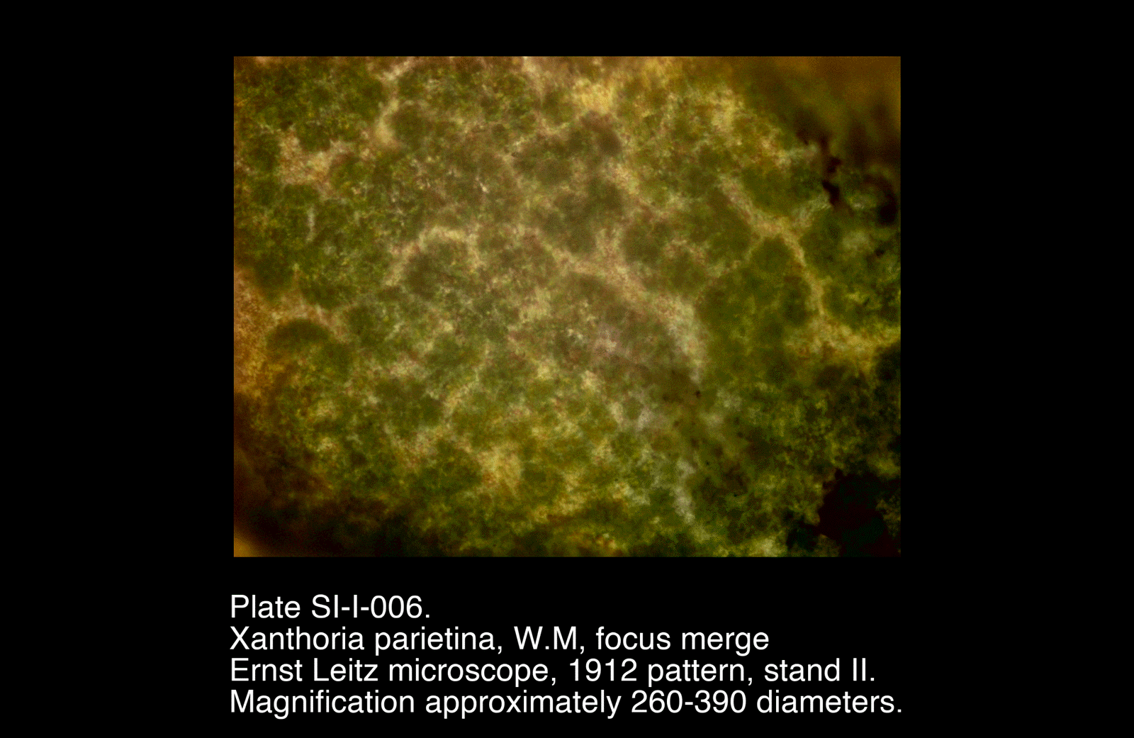
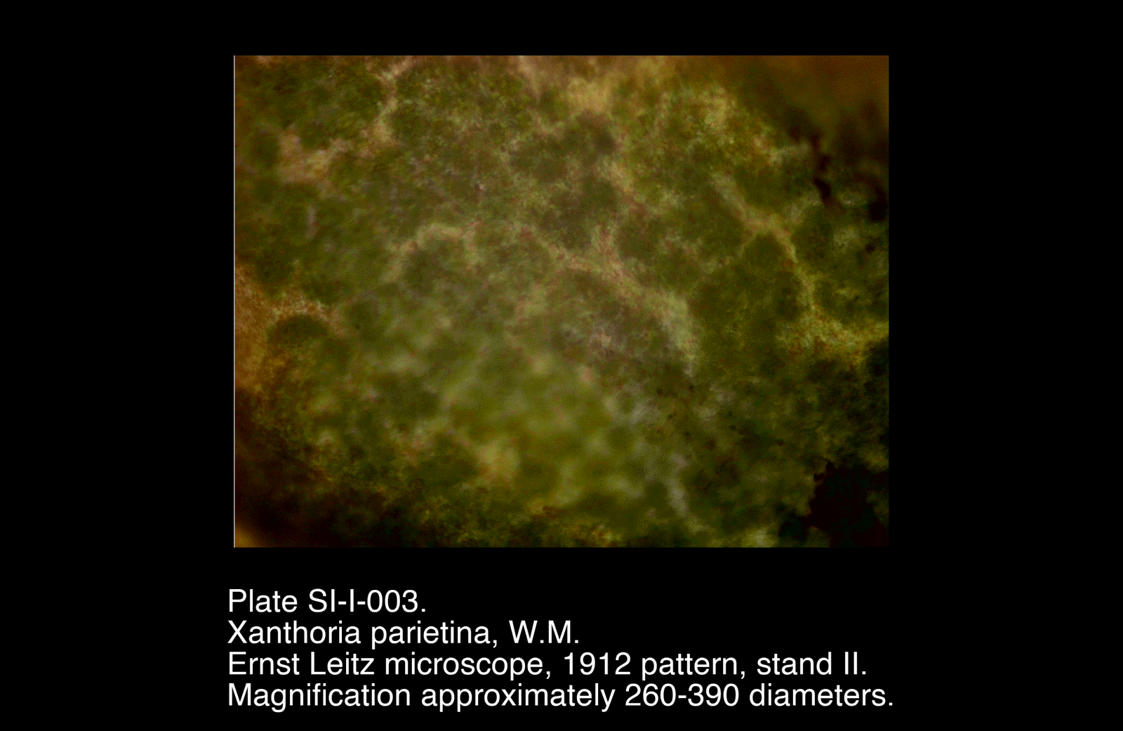
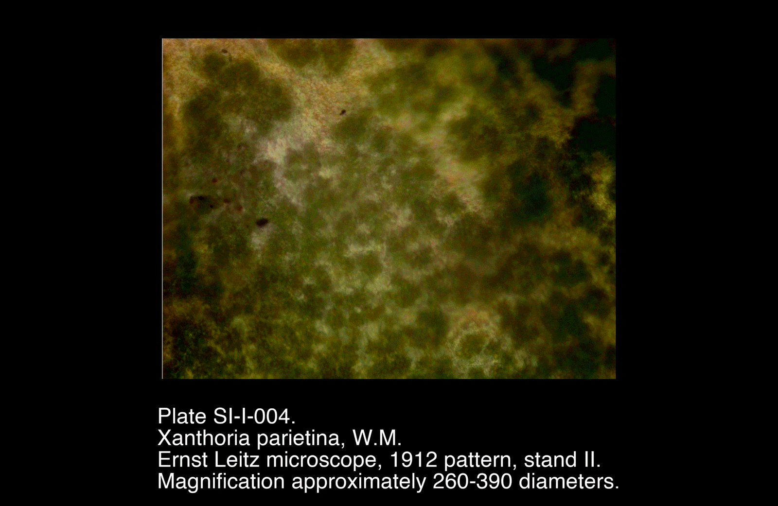
- Internal view shows a mottled / reticulate texture, not organised into the distinct cellular pattern expected in a higher plant epidermis
- Pale branching regions and darker patches are visible through the thallus
- No vascular organisation or differentiated supporting tissues observed
- Focus-merged plates provide improved definition of the surface and internal texture compared with the preliminary images
Plates
Selected Plates
/Xanthoria%20parietina,%20W.M,%20focus%20merge)
/Xanthoria%20parietina,%20W.M,%20focus%20merge)
Plates
Selected Plates
These plates show the clearest overall view of the lobed margin and the mottled internal texture of the thallus.
Preliminary Plates
/Xanthoria%20parietina,%20W.M.)
/Xanthoria%20parietina,%20W.M.)

/Xanthoria%20parietina,%20W.M.)
/Xanthoria%20parietina,%20W.M.)
Preliminary Plates
Earlier views were useful in establishing the general form of the thallus and in identifying the most informative regions for later imaging.
Interpretation
The structure seen in Xanthoria parietina differs fundamentally from that of a higher plant organ. Rather than showing discrete tissues specialised for support, conduction, and growth in the vascular sense, the lichen thallus appears as a composite body organised chiefly for surface exposure and exchange.
Surface Form
The lobed and expanding margin increases exposed surface area and allows the thallus to spread across the substrate. This growth form is characteristic of a foliose lichen and is well suited to interception of light, moisture, and dissolved atmospheric nutrients.
The uneven outline and gently folded surface may also help create small local variations in exposure and wetting.
Internal Organisation
The mottled and reticulate appearance suggests a body composed of interwoven fungal tissue with associated photobiont-containing regions, rather than the regular cellular fabric of a plant leaf or stem.
Although the present images do not fully resolve individual algal cells with certainty, the plates do suggest a heterogeneous internal arrangement, with lighter and darker regions corresponding to differences in density, texture, or composition within the thallus.
Functional Interpretation
The thallus may be understood as a surface-oriented symbiotic structure:
- Broadly expanded for exposure
- Thin enough to permit exchange across much of its body
- Lacking specialised conducting bundles
- Organised for persistence under alternating wet and dry conditions
This is consistent with the ecological habit of Xanthoria parietina, which thrives on exposed surfaces where light is abundant and hydration is intermittent.
Comparison with Higher Plant Material
Unlike the stems examined elsewhere in this series, Xanthoria parietina shows:
- No vascular bundles
- No obvious epidermis / cortex / pith differentiation in the vascular-plant sense
- No mechanically specialised outer ring for self-support
Instead, its organisation is that of a thallus, not an axis-bearing plant organ. Its structure reflects occupation of a surface rather than support of an upright body plan.
Remarks
- The focus-merged images (SI-I-005 and SI-I-006) are especially valuable in conveying the uneven relief of the specimen
- Whole mounting provided a useful first view of lichen structure, though a transverse section would likely reveal the layered organisation more clearly
- A future investigation comparing whole mount and sectioned material would be particularly worthwhile
- This specimen serves as a strong early example in Series I, illustrating a body plan centred on surface exchange rather than internal conduction