
Field Notes Journal Entry
Improving Hand Sectioning: Hedera helix Revisited
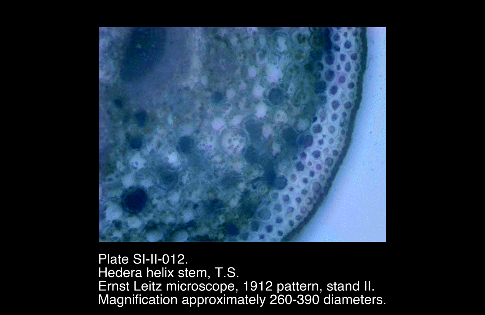
Revisiting Hedera helix stem sections using an improvised double-bladed sectioning tool, with noticeably improved consistency and structural clarity.
Revisiting earlier work on Hedera helix, I set out to improve the quality of transverse stem sections by refining the sectioning method.
In the initial experiment, sections were produced freehand using a single blade. This is barely good enough for even the most basic observation: sections were often too thick, uneven, and prone to tearing. This limited the clarity with which internal structures could be interpreted.
I wanted to keep a manual approach, for now, and while searching the web for improved sectioning techniques, I found a simple improvised sectioning tool based on a double-bladed design described on the Microscopy UK site - “How to make thin slices of specimens. And mount them.”.
The Sectioning Tool
The tool consists of:
- Two half-height, single-edged razor blades, carefully aligned in parallel
- Electrical tape to bind them together
- Bulldog clips to maintain rigidity and keep the two blades in place while cutting
This produces a double cutting edge which helps regulate the eveness and thickness of the resulting section.
Technique
Sections were taken by drawing the blade smoothly across the specimen rather than pressing downward. Multiple sections were cut in succession, and the thinnest were selected for mounting.
As noted in the article, I found that the best sections often collected between the two blades and could be retrieved with a fine brush once the blades were gently separated.
Observations
The improvement over the earlier freehand method was very marked, with sections being:
- Thinner and more consistent
- Cleaner at the outer boundary (epidermis and cortex)
- Less affected by tearing and compression
- Clearer in the vascular region
Most importantly, they moved from being indicative to being interpretable. In the case of Hedera helix, vascular structure could be followed with much greater confidence than before.